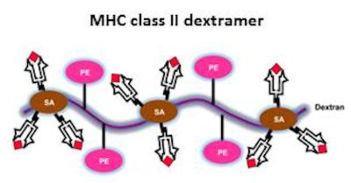
Major histocompatibility complex (MHC) class II tetramers and dextramers
Detection of antigen-specific CD4 T cells by flow cytometry
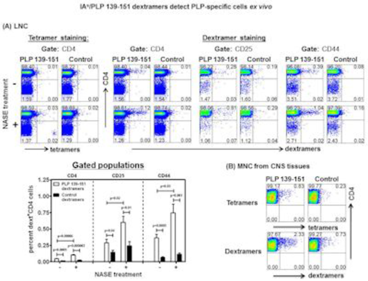
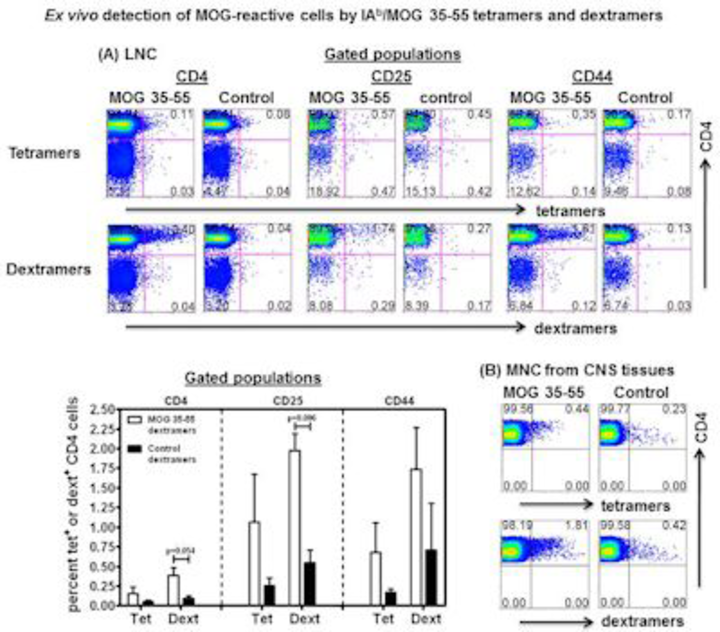
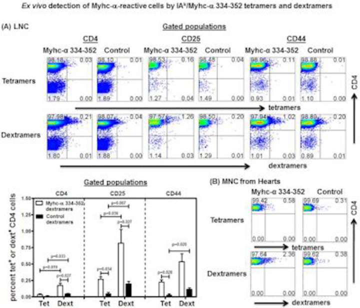
To determine the precursor frequencies and functionalities of antigen-specific, autoreactive CD4 T cells in various autoimmune disease models, we have generated a series of MHC class II tetramers and/or dextramers as noted below.
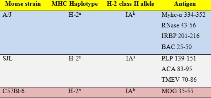
Abbreviations: Myhc, cardiac myosin heavy chain; RNase, ribonuclease; IRBP, interphotoreceptor retinoid-binding protein; BAC, Bacillus infantis; PLP, myelin proteolipid protein; ACA, Acanthamoeba castellanii; TMEV, Theiler’s murine encephalomyelitis virus; MOG, myelin oligodendrocyte glycoprotein.
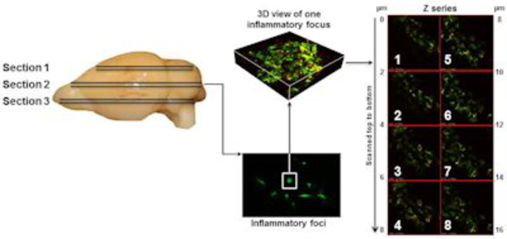
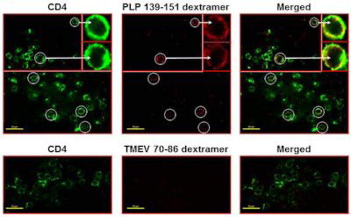
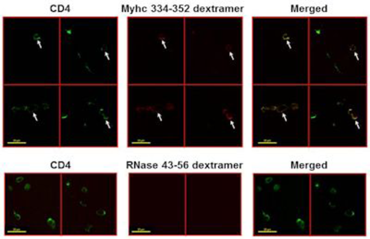
Detection of antigen-specific CD4 T cells in situ by confocal microscopy
We recently established a comprehensive method that permits us to enumerate the frequencies of antigen-specific, autoreactive CD4 T cells in situ in fresh tissues by direct staining without having to amplify the fluorescent signals, an approach commonly employed with conventional MHC tetramers. In particular, staining with dextramers is a one-step reaction, and the whole procedure, from tissue sectioning to staining, can be finished in less than a day.
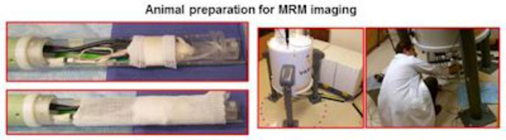
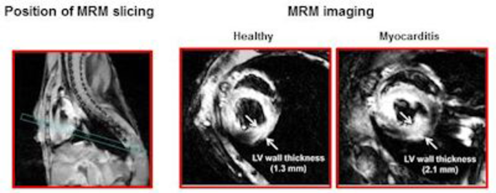
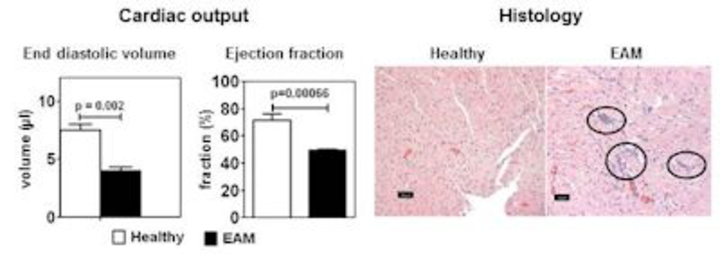
Magnetic resonance microscopy (MRM) imaging of heart in the mouse
To evaluate cardiac abnormalities in animals affected with myocarditis non-invasively, we have established a MRM imaging technique using a 9.4 T (400 MHz) 89 mm vertical core bore scanner equipped with a 4 cm millipede radio-frequency imaging probe and 100 G/cm triple axis gradients. The technique allows us to ascertain the thickness of the ventricular wall and functional changes in inflamed hearts, such as end diastolic and ejection fractions. Thus, MRM offers an advantage of assessing the progression and regression of myocardial injuries in diseases caused by infectious agents such as coxsackievirus B3, as well as response to therapies.